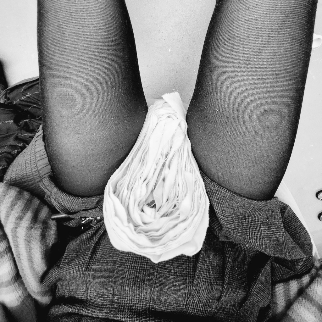
Contemporary visual arts practices that deal with defining the ambivalence between membrane and skin present as a consequence not only of something as systematic as semantics, but also as an exploration of the fragmentation pertaining to the organisation of surfaces that interact with the interior and exterior spaces.
To look inside the skin involves the piercing of itself. I want to explore the interplay of opening and enfolding.
discuss technicality of weaving – include artists – georgina-maximle – skin and stiching
MELISSA DUBBIN & AARON S. DAVIDSON // Delay Lines
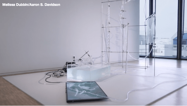
‘The story of our embryonic transformation is one that takes place in the amnion. We have no memory of this place, but our body remembers. Our body retains vestigial features of our time as fish. Tiny folds on our ears are the remnants of our gills, our inner ears one of the most ancient parts of our body.’
The installation pertains to the creation of artificial formations of life through computing software and the environments that robotics create for humans. The artists choose to describe the work from a very humanistic stance, they mention the amnion which is a thin membrane forming a closed sac about the embryo or fetus of a reptile, bird, or mammal and containing a watery fluid in which the embryo or fetus is immersed. In this way the amnion is the skin in which life is formed and nurtured within, a vessel containing sentient life.
2019
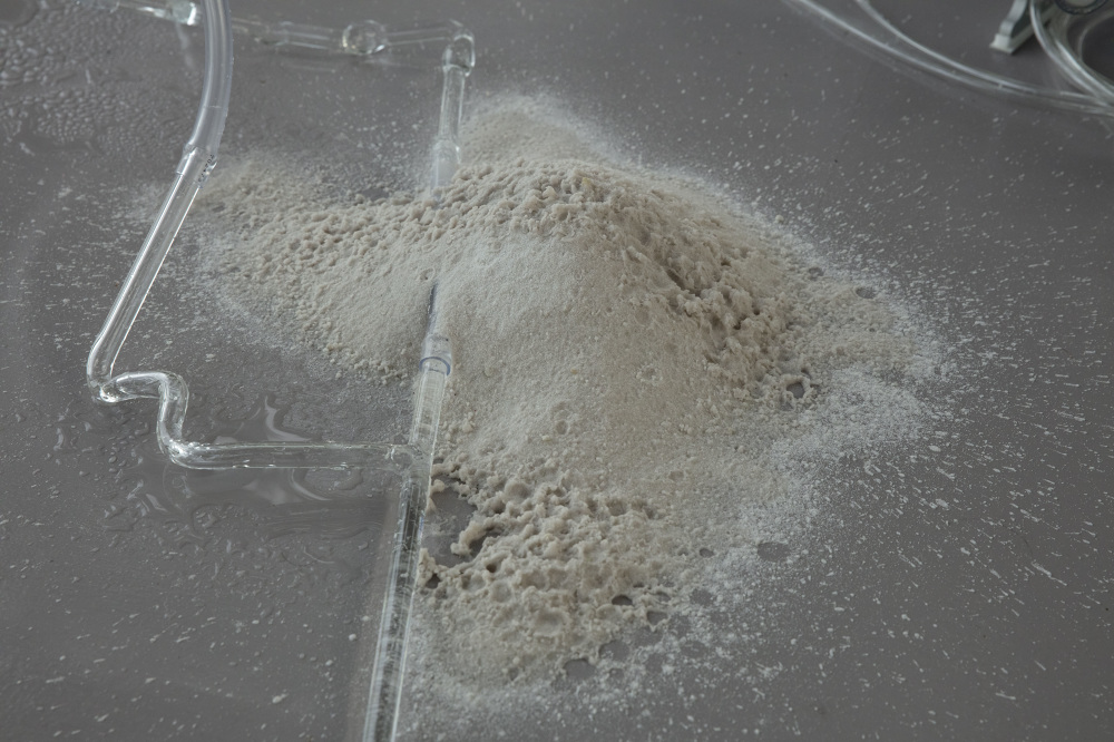
DETAIL // Water from subterranean Asahi River, borosilicate glass, overclocked water-cooled computer, soft robot manta, silicon boules, temperature sensors, micro-controllers, air compressor, air control system, simulated environment, monitor, metal, plastic and power supplies.
2.5 x 2.5 x 4 meters
Collection of Ishikawa Foundation, Okayama
Since Embryonic Vessels are systems for potential development, I have considered the shape and form of medical illustrations pertaining the the female reproductive system.
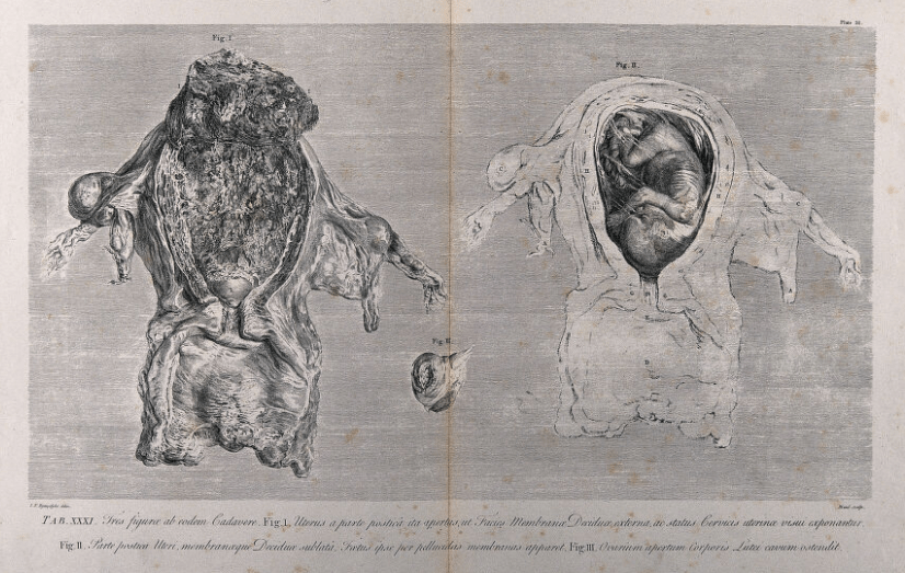
Rymsdyk, Jan van, active 1750-1788.
membrane reproduction / drawing series

Leave a comment